
An In-Depth Look at Ultrasounds
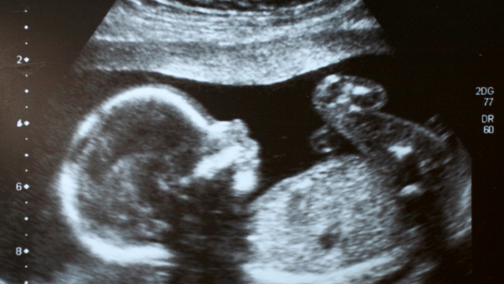
Pregnancy ultrasounds offer a window into the womb to monitor the development and well-being of the fetus. These non-invasive scans use high-frequency sound waves to create images of the baby and the uterus, providing crucial information that supports the health of both mother and child throughout pregnancy.
Ultrasound technology is fascinating as it transforms sound waves into visual data without the use of radiation, making it a safe choice for examining developing fetuses.
When the ultrasound probe, or transducer, is placed against the mother’s abdomen or inserted vaginally, it emits sound waves that bounce off the baby’s and other tissues’ structures. These echoes are then converted back into images by the ultrasound machine, revealing the hidden world of the developing baby.
The Purpose of Ultrasounds Throughout Pregnancy
First Trimester
In the initial stages of pregnancy, ultrasounds serve multiple purposes. They confirm the presence of the pregnancy, estimate due dates, and check the fetal heartbeat, providing the first audible reassurance of the baby’s health. These early scans also examine the placenta’s position and assess the risk of miscarriage or ectopic pregnancies, where the embryo implants outside the uterus.
Second and Third Trimesters
As pregnancy progresses, the focus of ultrasounds shifts towards monitoring fetal growth, determining the baby’s position, and identifying the baby’s gender, if desired. These scans are pivotal in evaluating the health of the placenta and the levels of amniotic fluid and screening for potential congenital abnormalities. Ultrasounds also guide healthcare providers in performing other tests, ensuring the safety and health of the fetus.

Types of Pregnancy Ultrasounds
Understanding pregnancy ultrasounds reveals the depth of prenatal care available to expectant parents.
These advanced imaging techniques, each with its unique application and benefits, play a crucial role in ensuring the health and safety of both mother and fetus throughout the pregnancy journey. Learn more about the various types:
- Transvaginal Ultrasound
- Ideal for the initial stages of pregnancy, providing an unparalleled view by placing the probe directly in the vagina, closer to the uterus.
- Superior for detecting early pregnancy viability, cervical assessments, and examining the placenta’s position about the cervix.
- 3-D and 4-D Ultrasounds
- 3-D ultrasounds offer a detailed static view of the fetus, allowing for examining physical features and potential anomalies in a way traditional ultrasounds cannot.
- 4-D ultrasounds extend the capabilities of 3-D imaging by adding time, showcasing the fetus’s movements in real time, and enriching the prenatal bonding experience.
- Fetal Echocardiography
- Specialized ultrasound focused on the fetal heart, utilized when there’s a suspected cardiac issue or a family history of heart defects.
- Provides detailed images of the heart’s structure, rhythm, and function, crucial for diagnosing congenital heart conditions and planning postnatal care.
Preparing for Your Ultrasound
Expectant mothers can prepare for their ultrasound by staying hydrated, as a full bladder can enhance the clarity of the images.
It’s important to wear comfortable clothing and to be aware that the appointment can vary in length, depending on the type of ultrasound and the specific measurements needed.

Safety and Considerations
The safety of ultrasound technology is well-documented, with no radiation involved and a long history of safe use.
However, it’s important to use this technology judiciously, adhering to medical guidelines for timing and frequency to avoid unnecessary exposure.
What Happens During an Ultrasound?
The ultrasound procedure is a seamless and non-invasive method that offers a glimpse into the womb, providing expectant parents and healthcare providers with vital information about the fetus’s development and health.
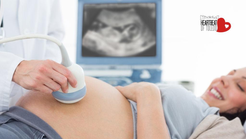
The process begins with the application of a conductive gel on the mother’s abdomen or, in the case of a transvaginal ultrasound, the insertion of the probe into the vagina. This gel acts as a medium that enhances the transmission of sound waves between the transducer and the skin, ensuring clear image quality. The sonographer, a trained ultrasound technician, plays a pivotal role throughout this procedure.
- Application of Conductive Gel: A water-based gel is liberally applied to the area of interest to reduce air pockets between the skin and the transducer, which could distort the sound waves and, consequently, the images produced.
- Transducer Manipulation: The technician maneuvers the transducer, a hand-held device, across the skin’s surface or inserts it into the vaginal canal, depending on the type of ultrasound being performed. This device emits high-frequency sound waves that penetrate the body and echo back upon encountering tissues, fluids, and bones.
- Image Capture and Analysis: As sound waves bounce back to the transducer, they are captured and translated into digital images displayed on a monitor. The sonographer adjusts the transducer’s position and angle to obtain images from different viewpoints, ensuring a comprehensive examination.
- Technician’s Expertise: The technician’s expertise is crucial in interpreting the live images. They identify structures, measure fetal growth, and assess developmental milestones, annotating these findings directly on the images for the healthcare provider’s review.
- Communication of Findings: While the sonographer may provide immediate observations, a radiologist or a specialized physician typically performs a detailed analysis and interpretation of the ultrasound images. The results are then communicated to the patient’s healthcare provider, who will discuss these findings with the patient, explaining their significance for the pregnancy’s progression and any further evaluations or interventions that may be necessary.

Understanding the Results
The images are reviewed after the ultrasound, and the results are communicated to the expectant parents, often during a follow-up consultation. This discussion can provide reassurance, offer insights into the baby’s health, and help plan for the remainder of the pregnancy and the delivery.
Recognizing the importance of these insights, Heartbeat of Toledo invites all expectant mothers to take advantage of our complimentary ultrasound services. Our dedicated team is committed to providing a supportive, informative experience, ensuring you receive the care and information necessary for a healthy pregnancy. Set up an appointment now!